 企业邮箱
企业邮箱


A Comparative Study of the Effects of Moist-Exposed Burn Oin
作者:Rong Xiang Xu 出版社:KARGER 发行日期:In 2004INTRODUCTION
Corneal epithelium consists of three types of cells at three different levels. They are basal cells in the deep layer, wing cells in the middle layer and squamous cells at the corneal surface. Microvillus in the superficial layer absorbs tears to form a tear membrane. Basement membrane behind the deep layer is located in the corneal Bowman membrane (anterior limiting lamina). Corneal epithelial layer is a natural barrier against micro-organism invasion. Once injured, it is prone to infection and shedding. Some corneal diseases such as dendritic keratitis, neuroparalytic keratitis, exfoliation of recurrent corneal epithelium, alkali burn, vesicular keratitis, etc., and surgeries such as scraping the corneal epithelium for intraocular examination and operation when performing cutting of the vitreous and repairing for retinal detachment, may injure corneal epithelium. Rapid and complete repair of corneal epithelial defects plays a very crucial role for the recovery of corneal physiological function and for good eyesight. Corneal physiological function contributes greatly to the promotion of deep corneal wound healing and formation of collagenous fiber [1]. In this study, rabbits with corneal epithelial defects of the same size were used and treated with MEBO, homologous serum, 0.5 % dexamethasone, 25,000 U/ml vitamin A. In addition, the rabbits wore soft corneal contact lens to observe the regenerating rate of corneal epithelia and to assess the therapeutic effects of different measures.
MATERIALS AND METHODS
Eighty-two healthy adult rabbits of either sex weighting 2~3 kg were anesthetized by 35% urethane 2.5 ml/kg intravenously and, if required, by inhalation of ether. Then 1 % dicaine was dropped twice for surface anaesthesia into the palpebral fissure which was expanded with eyelid retractor. The cornea center was constrictively marked at surface using an 8-mm corneal trephine and was stained with 1% fluorescein followed by rinsing with normal saline. Subsequent to the appearance of a circular mark, an 8-mm stained disk area of epithelial defect was created with a lancet scraping off the whole epithelium within cycle and by fluorescein staining.
The animals were then divided randomly into 6 groups, 12 in each. In group 1, the animals were treated with MEBO every 6 h. In group 2 to group 5, the animals were given eye drops with 0.5 % dexamethasone, 25,000 U/ml vitamin A, homologous serum and normal saline, respectively, every 2 h. In group 6, the animals wore soft corneal contact lens without any eye drops, whereby 6 eyes were removed of lens every 6 h for fluorescein staining and other 6 eyes stained 24 h later. In addition, 10 rabbits had their secretions cleaned away with cotton sticks soaked with normal saline and no eye drops were applied. Corneas were removed at 0, 2, 4, 6, 12, 18, 24, 30, 36, 48 h for microscopic observation on corneal epithelial healing [2].
RESULTS DISCUSSION The experiment revealed us the two periods for epithelial healing. At 2 h postsurgery, the defects did not decrease, instead, some increased from 8 to 8.1~8.5 mm, which may have resulted from the contraction of tissues at the wound edge and the shedding of injured cells. It was not until 6 h postsurgery that epithelial defects began to diminish progressively until complete wound healing was achieved. Systemic or local administration and either intra- or extra-ocular agents may be factors contributive to the successful repairing process of corneal epithelial defects from the latent period till the end of healing period. By means of smearing, eye drops or wearing of corneal contact lens, the study investigated various factors relative to epithelial healing in order to verify the therapeutic effects of methods and drugs. MEBO is an ointment containing sesame oil, beeswax and other active ingredients extracted from plants such as Cortex Phellodendri, Radix Scutellariae. It is highly effective in the treatment of burns, wounds and ulcers and its advantages include superior analgesic effects, anti-inflammation, infection prevention and minimal scar formation post healing [3]. Although recent reports prove MEBO is significantly superior to other therapies in treating various burns, wounds and ulcers [4], there is no comparative study on the management of eye injury reported. Therefore, we conducted a preliminary comparative study on fibronectin (FN) and MEBO in the treatment of experimental corneal alkali burns in rabbits, which verified that MEBO was remarkably effective in the treatment of burns on avascular corneal tissue [2, 5]. Further, we performed this study to investigate the effect of MEBO on repairing and healing of corneal defect and the roles of other drugs as well. The results verified that MEBO is obviously superior to other medications excepting for homologous serum in promoting wound healing while no obvious corneal macula was formed posthealing under slit-lamp microscope examination. The pharmacological mechanisms of MEBO with respect to its role of promoting healing of corneal wounds also became understood as follows: (1) Composed of a macromolecular sticky base, MEBO has an affinity to tissue protein at the wound site. The application of MEBO to the wound may serve as a bridge to directly stimulate and induce cell division and migration in an orderly manner that promotes wound healing. (2) MEBO contains various nutrients necessary for wound healing. For example, glucose is an obvious energy source. Vitamins and organic acids, which are related to the maintenance of tissue metabolism and proliferation of connective tissues, may directly support local nutritional needs, thereby preventing scar formation [6]. Zinc and enzymes may accelerate epithelial repair [7]. Protein as the basic element of the cell membrane may support the growth, differentiation and regulation of cells. (3) MEBO promotes the formation of a unique, integrated automatic drainage circulation system which corrects for local dysfunctional metabolism and circulation resulting from injury. The base ingredients contained in MEBO absorb metabolic products from the wound and then transport them to the outer layer of the ointment. Meanwhile, active ingredients of the ointment continuously penetrate into the wound to renew the supply of ingredients necessary for tissue repair. The automatic microparticle transportation and processing of emulsification and dispersing are considered as the main measure for the treatment of injured avascular tissue [8]. (4) Obaculactone contained in MEBO offers properties of anti-inflammation, detumescence, analgesic effects, and enhancing local immunity and controlling infection. Many reports demonstrate the presence of FN in plasma. FN is a macromolecular glycoprotein that is the conjunctive media between cell and extracellular fibers and matrix. It adheres to collagen, polysaccharid protein, and receptors on the cell surface and serves as an intercellular bridge of epithelial cells. It has some correlation with cytoskeleton structures, e.g. microfilament, actin, to induce the migration of the cell. FN plays an important role in the firm adhesion between migrating epithelial cells and wound surface of corneal epithelial defect. Eye chemical burns can be treated with eye drops or subconjunctival injection composed of autoblood. Autoblood is beneficial because it contains macroglobulins that inhibit collagenase, release fibrinolysin (which may reduce symblepharon, thereby promoting the recovery of the blood vessel net around the cornea as well as restoring sensation in the injured corneal), improve corneal nutrition and promote tissue regeneration. Our previous study has verified that FN can speed up the migration of corneal epithelial cells [2]. Observation on the rabbit corneal epithelial healing rate after application of homologous serum in this study has also suggested an obvious effect of serum in promoting the migration of corneal epithelial cells. However, even serum offers less benefit than does the application of MEBO. Various reports can be found about the role of corticosteroid in corneal wound healing. It is believed that long-term administration of corticosteroid at high concentration may retard epithelial regeneration and that stromal wound healing though the mechanism remains unclear. On the other hand, the use of corticosteroid immediately after corneal burns may have good anti-inflammatory effects and reduce occurrence of ulcers and neogenetic vascularization [9]. Others report that corticosteroids may inhibit conjunctival cells from migrating towards the corneal surface without impairing the reformation of corneal epithelium [10]. In this study, eye drops of 0.5 % dexamethasone did not inhibit the migration of corneal epithelial cells and wound healing. On the contrary, it significantly promoted the healing. Therefore, it is feasible to drop 0.5 % dexamethasone on simple corneal epithelial defects to control inflammation and promote healing, though even this modality, due to adverse effects, is far inferior to the effects achieved by MEBO. Vitamin A plays a role in promoting epithelial growth and maintaining epithelial normal functions. Many reports have confirmed vitamin A contributing to promoting wound healing, but some researchers reported that vitamin A failed in promoting epithelial regeneration. The use of 25,000 U/ml vitamin A in this study revealed little effect on corneal epithelial healing. Corneal epithelium, when wearing corneal contact lenses made of polymethyl methacrylate (PMMA), is provided with nutriments and oxygen necessary for metabolism which derive primarily from tears penetrating between the lens and cornea. Eyelid pressure when eyes are open produces a positive pressure behind the lens to discharge tears and the negative pressure produced behind the lens after winking allows the entrance of the tears to float the lens. Such a process goes in cycles to achieve the exchange and renewal of tears. Subsequent to corneal epithelial injury, the ability of local metabolism decreases and wearing lenses further impairs oxygen absorption of the cornea, which is unfavorable to wound healing. Though some reports suggested that wearing corneal contact lens might be used for treating corneal ulcer, the observation in this study verified that such management impaired the corneal wound-healing process and healing rate was much lower than those in other groups. We conclude that careful attention should be taken to avoid harming patients through the application of corneal contact lens in the treatment of corneal injury. REFERENCES
The observation record showed the following mean healing time: 48.6 h in the normal saline group, 47.36 h in the vitamin A group, 45.18 h in the homologous serum group, 42.78 h in the MEBO group, 46.5 h in the dexamethasone group and 65.3 h in contact lens group. The results revealed that there was no significant difference of average healing rate between groups 1 and 4, as well as groups 2 and 4, groups 2 and 3, groups 3 and 5. However, there were statistical differences between other each two groups. MEBO, homologous serum, vitamin A, and dexamethasone had superior healing effects compared with normal saline, while wearing of contact lens retarded the healing of the defect. Beside homologous serum, MEBO is remarkably superior to other drugs in promoting corneal epithelial healing rate (Table 55).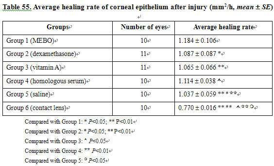
Cornea has sources of nutrition from the vascular net in corneal limbus, tears and aqueous humor. Corneal epithelium is a functional barrier between tear membrane and intraocular tissues through which the fluid output from the stroma is regulated in order to keep the stroma in a normal hydration. A corneal epithelial defect caused by injury or scraping off can repair rapidly in two stages. The latent period comes first, with a mean time of 5.50.3 h during which extensive cellular and subcellular changes at the wound edges are expressed by desquamation of superficial cells, by loss of columnar appearance of basal cells, by damage on hemidesmosome link of the basement membrane as well as by formation of a cell process,indicating that residual viable epithelial cells are transforming into functional cells. A healing period is followed afterwards when epithelial cells around the wound migrate towards the center at a constant pace without mitochysis. This process starts from the uninjured epithelial cells adjacent to wound edge. We see basal cells in particular being enlarged and flatted with pseudopodium migrating towards the center and becoming the migratory edge of monolayer cells, which is followed by two or more layers of epithelial cells. The migration stops as wounds completely close and a firm hemidesmosome adhesion to the basement membrane is re-formed. At this point, mitochysis begins and mature corneal stratified epithelium is finalized.
1. Smolin G, et al: Tretinoin and corneal epithelial wound healing. Arch Ophthalmol 1979; 97: 545.
2. Huang QS, et al: A comparative study of fibronectin and MEBO in the treatment of experimental corneal alkali burn in rabbits. Chin J Burns Wounds Surface Ulcers 1995; 7: 18.
3. Xu RX: The medicine of burn and ulcer: A general introduction. Chin J Burns Wounds Surface Ulcers 1989; 1: 11.
4. Xu RX: The principle of burn wound treatment. Chin J Burn Wounds Surface Ulcers 1992; 4: 8.
5. Huang QS, et al: A dynamic study on MEBO repairing corneal endothelium alkali burn in rabbit. Proceedings of the Fourth National Conference on Burns, Wounds and Ulcers, Beijing, 1995.
6. Huang QS, et al: Clinical observation of herpes simplex corneal ulcers treated by combined MEBO and PIC (25 cases report). Chin J Burns Wounds Surface Ulcers 1993; 5: 21.
7. Morley TE, et al: Zinc deficiency chronic starvation and hypothalamic pituitary thyroid function. Am J Clin Nutr 1980; 33: 176.
8. Du HE: A preliminary introduction on relationship between the biopharmacy factors and treatment of MEBO. Chin J Burns Wounds Surface Ulcers 1995; 7: 8.
9. Woost PG, et al: Effect of growth factors with Dexamethasone on healing of rabbit corneal stromal incisions. Exp Eye Res 1985; 40: 47.
10. Srinirasan BD: Corneal re-epithelialization and anti-inflammatory agens. J Am Ophth Soc 1982; 80: 756.