 企业邮箱
企业邮箱


Experimental research on the Mechanism of the Anti-infection
作者:Rong Xiang Xu 出版社:KARGER 发行日期:In 2004INTRODUCTION
Many basic studies and clinical research have proved that BRT with MEBT/MEBO has beneficial effects of anti-infection, promoting burn wound healing and reducing scar formation to name but a few of its attributes. However, the mechanisms of these effects of MEBO remain a bit mysterious. The following study was designed to elucidate the anti-infection effect mechanism of BRT with MEBT/MEBO.
MATERIALS AND METHOD
Apparatus and Reagents:
Fully automatic enzyme labeling analyzer, Multiskan, MS; AC-920 hemocyte analyzer; MEBO; RPMI-1640, Gibco, USA; Con A, supplied from Guangzhou Medical Institute; MTT supplied from Fluka; Pathogenic O111B4 of Escherichia coli supplied from Binzhou Medical College.
Animals and Methods:
Thirty-two BALB/C mice, 8 weeks old, females, weighing 17~19 g were supplied from Beijing Medical University. Twenty-eight Kunming mice of both sexes weighing 21~24 g, 18 rabbits weighing 2~2.5 kg and 4 guinea pigs weighing 250~300 g were supplied from Binzhou Medical College.
The animals were divided randomly into a control group and a MEBO treatment group. In the MEBO treatment group, mice were depilated (2 2 cm) on their backs and smeared with MEBO. Guinea pigs were depilated (3 2.5 cm) on their backs and smeared with MEBO. MEBO was applied 2 times a day. Biopsy of skin tissue was done on day 9 to determine the activity of interleukin-1 (IL-1), and was stained with hematoxylin and eosin (HE) for histopathological observation.
Examination Indexes:
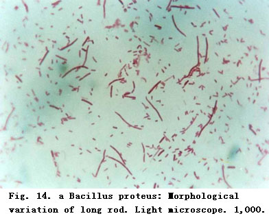
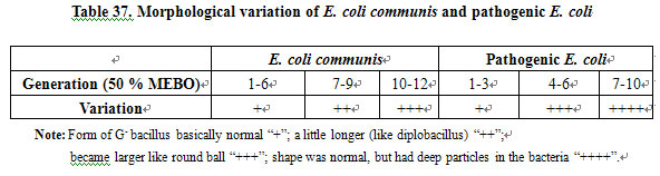
Morphological Variation of Pathogenic E. coli (O111B4). The bacteria were cultivated on culture medium containing a given concentration of MEBO [1]. Each generation of the bacteria was stained using the G staining method. Staining reaction and morphological variation of the bacteria were observed. The results were compared with that of the E. coli communis cultured on the same kind of medium containing MEBO.
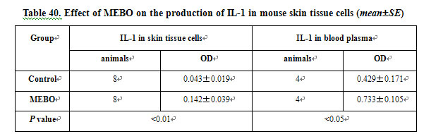
Determination of IL-1 Level. MTT was used as a substrate. Since living cells containing active succinate dehydrogenase can reduce yellow-colored MTT to form violet or blue-colored formazam particles, the particles were dissolved by adding isopropyl alcohol hydrochloride [2]. The OD value was determined by colorimetry. The amount of formazam was proportional to the number of living cells. Determination was performed according to the modified methods described previously[2]. Mice was killed under sterile conditions, the thymus was removed, cut and screened. After being centrifuged, a suspension with 107/ml of cells containing 2μg/ml of Con A was prepared. On a 24-pore plate, we added to each pore 0.5 ml of this suspension and then added mice skin tissue suspension and blood plasma at 0.5 ml/pore. Two more control groups, one with 1640-nutrient fluid (thymocytes plus nutrients) and the other with Con A plus thymocytes were set. The 24-pore plate was placed into an incubator at 37C, 5 % CO2 for 68 h. From each pore, 0.5 ml of the supernatant was drawn away, then 0.1 ml of 5μg/ml MTT solution was added to each pore and incubated again for 4 h. 0.5 ml of isopropyl alcohol hydrochloride was added to each pore and then the plate was shaken to dissolve the particles. Supernatant from each pore was drawn and added to a 96-pore plate, 0.2 ml/pore. The plate was placed into a fully automatic enzyme-labeling analyzer to determine the OD value, reflecting the level of IL-1 in skin tissue cells and blood plasma.
Effect of MEBO on Body Temperature of the Rabbit. The body temperature of the rabbit was taken prior to application of MEBO. Then, an area of 6.06.5 cm was depilated on the back of the animal and MEBO was applied twice a day while body temperature was recorded simultaneously. This was done for a total of six measurements. Changes of body temperature were recorded.
Effect of MEBO on the Classification of Mice Leukocytes. Every mouse in the MEBO and control groups had a 0.2-ml blood sample taken by enucleation of the eyeball. The blood was added to 10 ml of the reagent solution and then examined using an AC-920 hemocyte analyzer.
Assay of Humoral Immunologic Function. A quantitative hemolysis spectrophotometry (QHS) method was used to determine the amount of hemoglobin released after hemolysis of RBC mediated by antibody-forming cells [3]. This amount (expressed as OD value) reflected the amount of antibody-forming cells in mice, thus indicating the humoral immunologic function of the mice.
Assay of Cellular Immunologic Function: Blood samples were taken from mice tails in the MEBO and the control groups, respectively, then smeared and stained according to the α-naphthalene acetate esterase (ANAE) method. These were then examined under the microscope. 100 lymphocytes were observed randomly. The percentage of ANAE-positive lymphocytes reflects the cellular immunologic function of the body.
Histological Changes of Mice Skin after Treatment with MEBO: 0.5-cm2 skin tissue samples of mice taken from depilated areas of normal skin and skin treated with MEBO were fixed, embedded, stained with hematoxylin and eosin and then observed under the light microscope.
RESULTS
1) Anti-infection Effect of MEBO
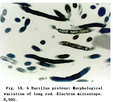
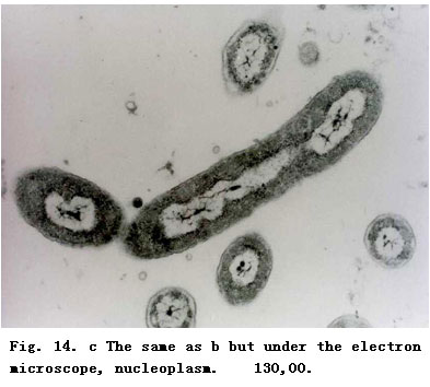
Morphological Variation of Bacteria. The results of morphological variation of E. coli communis and pathogenic E. coli cultured in medium with MEBO are shown in Figure 14 and Table 37. These reveal that MEBO acts to induce the variation of both E. coli communis (which is common in burns wounds), and pathogenic E. coli. However, the variation of different bacteria might occur at different times.
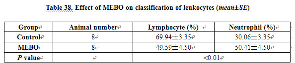
Effect of MEBO on the Classification of Circulating Leukocytes: Table 38 shows that the amount of neutrophil in blood increased after MEBO treatment.
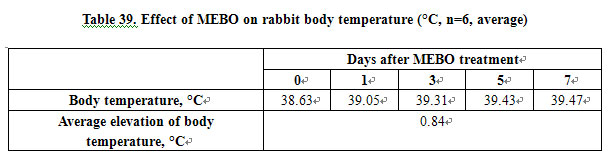
Effect of MEBO on Rabbit Body Temperature. 75% of the rabbits had an increase in body temperature after MEBO treatment. On day 7, the average elevation of body temperature was 0.84°C (Table 39).
MEBO Promoted Proliferation of Skin Cells and Cells at the Margin of the Sebaceous Gland. In this study, we found that the number of skin basal cells in the division phase was increased, and the number of juvenile flat cells at the margin of the sebaceous gland was also increased in the MEBO group. This finding indicates that the metabolism of the cells was vigorous.
4. Effect of MEBO on the Specific Immunologic Function of Mice
Table 41 shows that MEBO did not affect the cellular and humoral immunologic functions. 
CONCLUSION
The results suggested that: 1) MEBO can prevent infection; 2) the wound-healing benefit and the reduction of scar formation in the MEBO group were related to its effect of increasing the production of IL-1 by skin cells.
Table 41. Effect of MEBO on specific immunologic function of mice (meanSE)
DISCUSSION
Clinical practice has strongly proved that MEBO has anti-infection, pain-killing, wound-healing-promoting and scar-forming-reducing effects. Based on our study, we discussed the mechanisms of its actions as the following.
1) Anti-Infection Effect of MEBO: MEBO has potent ability to control wound infection and keep the wound moist but not macerated. The active ingredients in MEBO ointment and its unique dosage delivery system create an environment hostile to bacterial growth. In culture medium containing MEBO, morphological structural and physiological variations of the bacteria occurred. MEBO affected the synthesis of the components for formation of a bacteria wall and inhibited related enzymes. Also, the synthesis of DNA was inhibited and the bacteria proliferation rate was decreased. Deep pigments were found in the bacteria, this indicated that the bacteria were in a stable phase of proliferation, during which a high level of glycogen, lipid, etc. was stored in the bacteria.
MEBO induced morphological and physiological variations in bacteria while influencing the production of plasma coagulase of Staphylococcus aureus [4]. Bacterial pathogenicity is related to the bacterial wall component and thus MEBO reduced the pathogenicity of the bacteria. The bacterial variation characteristics were varied in different species of bacteria and at different MEBO concentrations. The initiation time of the variation was not standard.
In clinical care, we found that after treatment with MEBO, the body temperature of burn patients might rise by 1-1.5°C during the initial stage. At 3 h after application of MEBO, the temperature of burn patients with superficial second-degree wounds rose. In this study, the body temperature of rabbits increased after application of MEBO, and rose by 0.84°C on day 7. Furthermore, MEBO induced the production of IL-1, from IL-1-delivering skin cells, such as epidemic cells, keratinocyte, and Langerhans cells. In the early 1940s, people recognized that certain extracts from acute inflammatory foci could cause fever after injection into the body. This type of inflammatory substance is called endogenous pyrogen (EP). In 1979, purified EP was first proved to have IL-1 activity, and EP and IL-1 were considered as the same molecule [5]. MEBO stimulates local skin cells to produce IL-1, which in turn was absorbed into the systemic circulation thereby effecting the temperature-regulating center leading to elevated body temperature.
The effect of fever on body resistance of mammals is not clear yet. Some researchers think that fever may promote the immunity of the host. When body temperature is raised, but still <41°C, the phagocytic power of most phagocytes is enhanced. We also found that MEBO can promote phagocytic power of abdominal cavity macrophages in mice [6]. In this study, the quantity of neutrophils in blood circulation was significantly increased after treated with MEBO. Bone marrow stimulated by IL-1 may account for this interesting finding.
2). MEBO Promotes Wound Healing and Reduces Scar Formation:
Clinical data revealed that after treatment with MEBO, patients with superficial or deep second-degree burns wounds healed with full epithelization; superficial third-degree burns wound healed with a mild scarring that appeared smooth and soft. In this study, we observed that the production of IL-1 was increased in mice skin and sub-dermal tissues after treatment with MEBO. The difference between the MEBO group and the control group was very significant. Besides macrophages, many other tissue cells when stimulated can produce IL-1 in 1 h [9]. IL-1, IL-8 and tumor necrosis factor (TNF) are cellular factors which are capable of activating and inducing differentiation of T and B lymphocytes, enhancing the activities of monocytes, NK cells and killer cells, thus stimulating lysosomal enzyme activity and phagocytic activity of neutrophils.
Recently, both animal experiments and clinical practice proved that IL-1 does induce a series of pathophysiological changes. These changes are similar to the host’s response to infection [6], indicating that IL-1 is an important regulatory factor of the body as regards inflammation and immunologic reaction. We must differentiate between the local effect of IL-1 and the effect of high levels of IL-1 systemically. These are two totally different concepts [6]. In this study, the level of IL-1 in mouse skin and extracellular subdermal tissues in the MEBO treatment group were significantly different from those of the control group, as well as the IL-1 level in blood plasma. IL-1 is closely related to wound healing and we know that wound exudate typically contains IL-1. IL-1 promotes proliferation of fibroblast and secretion of collagenase [6]. The effect of IL-1 is complicated. It induces inflammation and fever while at the same time promoting wound healing. Inflammation induced by IL-1 is understood to be a kind of host defense reaction [7]. After mice were treated with MEBO, their skin basal cell in division stage increased. Juvenile cells observed around the sebaceous gland were very metabolically active. This proves that MEBO promotes wound healing.
3) Effect of MEBO on Specific Immune Function:
In this study, MEBO could increase the quantity of neutrophils in blood, while relatively decreasing the number of lymphocyte. In addition, MEBO did not affect the cellular and humoral immunologic function.
REFERENCES
1. Qu YY, et al: Experimental research on the anti-infective mechanism of MEBO. Chin J Burns Wounds Surface Ulcers 1996; 40: 19-23.
2. Mosmann T: Rapid colorimetric assay for cellular growth and survival: Application to proliferation and cytotoxicity assays. J Immunol Methods 1983; 65: 55.
3. Bi AH (ed): Medical Immunology. Tongji, Tongji Medical University, 1986, p7.
4. Yu H: Medical immunology. Beijing, People’s Health Publishing House, 1983, p 54.
5. Yang GZ (ed): Outline and Technology of Immunology and Bioengineering. Jilin, Jilin Science & Technology Press, 1991, pp2-15.
6. Chen WJ (ed): Molecular and Cell Biology of Blood. Beijing, Chinese Medical Science & Technology Press, 1993, vol 126-127, p 362.
7. Yang GZ, et al: Progress in genetic engineering and clinical immunology; in: Domestic and International Progress in Medical Sciences. Shanghai, Shanghai Institute of Medical Science & Technology Information, 1987, p124.